
A Case of Severe Adolescent Scoliosis Treated with Precise Planning and Robotic-Assisted Spine Surgery
A 12-year-old girl from North Karnataka was brought to SpineDRx after her parents noticed a gradual bend in her back and an unusual prominence around her ribs. Her mother first noticed a rib hump while bathing her, while her father observed that she was not sitting upright while doing her homework.
An initial evaluation by a local orthopaedic doctor, including an X-ray, revealed scoliosis, a condition in which the spine develops an abnormal sideways curvature.
Concerned about the progression of the deformity, the family travelled to Bangalore for a specialist spine consultation with Dr. Ramachandra Govindasamy at SpineDRx.
Patient Presentation
The patient was a 12-year-old girl with:
- Progressive spinal curvature
- Visible rib hump
- Postural changes while sitting
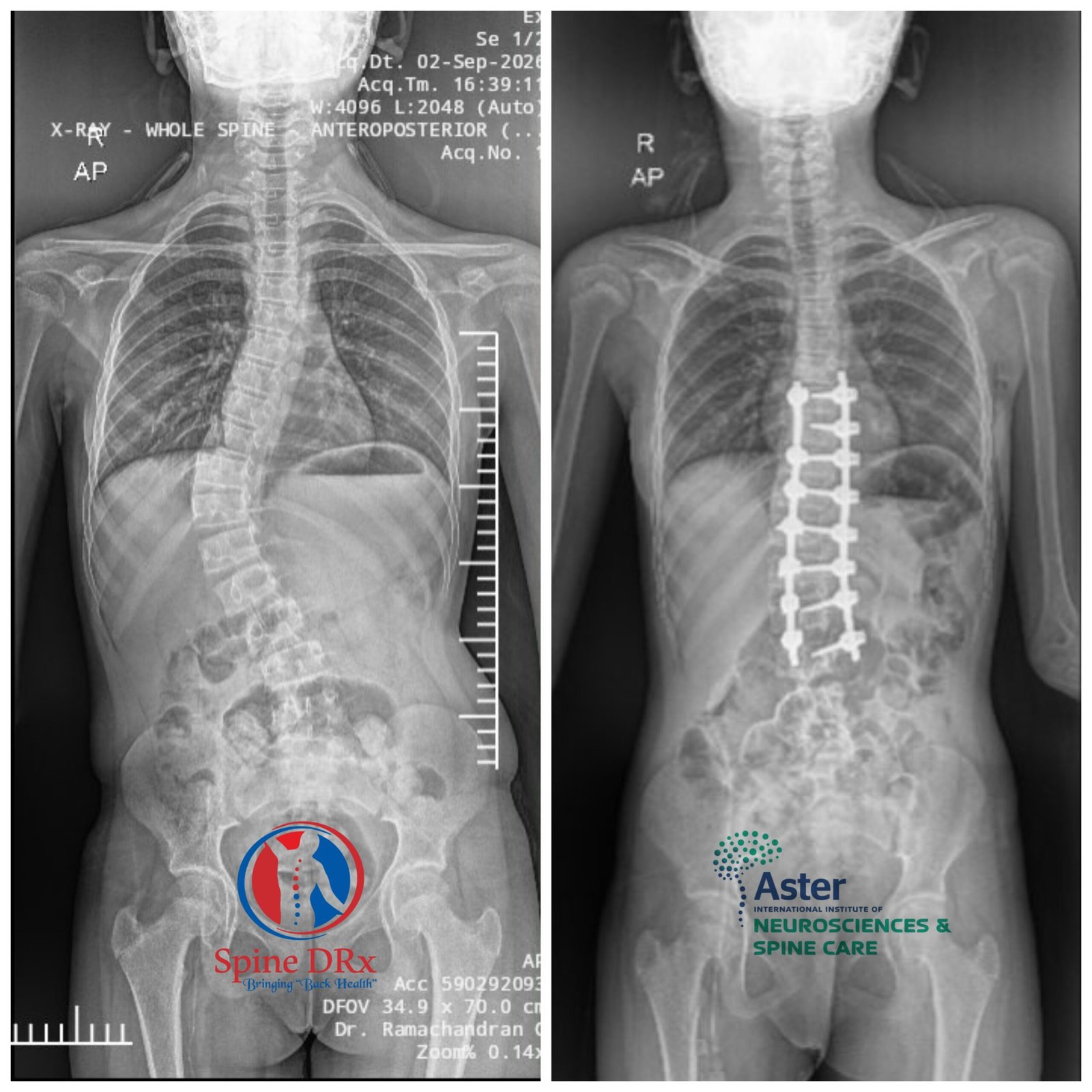
- A scoliosis curve measuring more than 50 degrees
- Skeletal maturity indicating that most of her growth had been completed
- Approximately three months since menarche
The family was prepared to consider different treatment options depending on the assessment, including bracing, growth-friendly procedures or corrective surgery.
Following a detailed clinical and radiological evaluation, it was determined that she was skeletally mature enough for definitive spinal fusion surgery.
Why Skeletal Maturity Was Important
In children with severe scoliosis, the patient's remaining growth is an important factor when deciding the appropriate treatment.
For younger children who still have significant growth remaining, particularly those with severe curves, growth-friendly techniques such as growing rods may sometimes be considered.
In this patient, however, her age, post-menarche status and radiological indicators of skeletal maturity suggested that she had reached a stage where a definitive fusion procedure could be considered.
This allowed the surgical team to plan a one-time corrective fusion procedure rather than a growth-friendly procedure requiring repeated lengthening or adjustments.
Detailed Pre-Operative Planning
One of the most important aspects of this case was the detailed assessment and planning before surgery.
Standing X-rays and side-bending X-rays were performed to understand the flexibility and characteristics of the spinal curvature.
The curve pattern was carefully analysed and identified as a Lenke 1AR curve, an important classification for determining the appropriate levels for correction and fusion.
An MRI was also performed to evaluate the spinal cord and rule out associated spinal cord abnormalities.
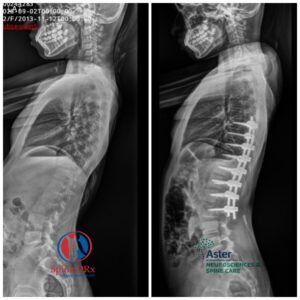
Based on the clinical and radiological findings, the surgical team planned a limited fusion from T7 to L2.
The goal was not simply to straighten the spine. The surgical plan also aimed to:
- Achieve substantial correction of the deformity
- Correct the rib prominence
- Preserve as much spinal mobility as possible
- Maintain appropriate balance between the thoracic and lumbar spine
- Allow the patient to continue activities such as sports and dancing
- Preserve forward bending ability
- Avoid unnecessary fusion of additional spinal levels
Robotic-Assisted Scoliosis Surgery
The corrective procedure was performed using robotic-assisted spine surgery, with intraoperative imaging and navigation technology supporting precise screw placement.
The surgical plan was created before the procedure, and the robotic system helped guide the placement of the screws according to the planned trajectory.
The procedure was performed with continuous neuromonitoring to monitor important neurological signals throughout surgery.
Intraoperative CT imaging and robotic navigation were used to assist the surgical team in achieving accurate implant positioning while protecting the surrounding neural structures.
90% Correction of the Spinal Curve
The pre-operative scoliosis curve measured more than 50 degrees.
Following the corrective procedure, the residual curve was approximately 5 degrees, representing an approximately 90% correction based on the measured angles.
The visible rib hump was also significantly corrected, resulting in a substantial improvement in the patient's overall spinal alignment and cosmetic appearance.
Because the curved spine was straightened, the patient also gained approximately one inch in height following surgery.
Early Mobilisation and Recovery
The patient's recovery following surgery was encouraging.
- Mobilised within approximately 3 hours
- Discharged in less than 2 days
- Returned to routine daily activities
- Able to attend school
- Able to bend and perform normal personal activities
- Able to manage daily hygiene and eating independently
The early recovery demonstrated the importance of careful surgical planning, appropriate patient selection, precise execution and structured post-operative care.
The Challenge Behind the Surgery
In severe scoliosis, achieving correction is not simply a matter of placing screws and rods and straightening the spine.
A major challenge is predicting how the remaining thoracic and lumbar spinal segments will respond to the correction.
The surgical team must determine:
- Which levels need to be included in the fusion
- How much correction is appropriate
- How to maintain overall spinal balance
- How to avoid creating secondary deformities
- How much spinal mobility can be preserved
- How to achieve correction without unnecessarily extending the fusion
This is particularly important in young patients, where preserving healthy, mobile spinal segments can influence their long-term function.
In this case, detailed pre-operative imaging and planning enabled the surgical team to select a limited fusion from T7 to L2, balancing correction with preservation of spinal mobility.
Technology Supporting Precision
The procedure was supported by advanced surgical technology, including:
Robotic Navigation:
Used to assist with accurate placement of spinal implants according to the pre-operative plan.
Intraoperative CT Imaging:
Provided detailed imaging during surgery to help confirm anatomy and implant positioning.
Neuromonitoring:
Continuous monitoring of neurological signals during the procedure provided an additional layer of safety.
The combination of clinical expertise, detailed planning, intraoperative imaging, robotic assistance and neuromonitoring helped the surgical team achieve the planned correction.
Outcome
The patient achieved an excellent radiological and clinical outcome following surgery.
The spinal curve improved from more than 50 degrees to approximately 5 degrees, with significant correction of the rib hump and improvement in overall appearance.
She was mobilised within hours, discharged in less than two days and returned to her normal daily activities.
Most importantly, the limited fusion strategy was planned with the aim of preserving useful spinal movement and allowing the young patient to continue activities she enjoys, including sports and dancing.
Key Takeaways from This Case
This case highlights the importance of early recognition, accurate classification and meticulous pre-operative planning in adolescent scoliosis.
For a young patient with a severe spinal curve, treatment decisions depend on several factors, including:
- Age
- Remaining skeletal growth
- Curve magnitude
- Curve flexibility
- Spinal balance
- Neurological status
- Overall health
- Functional requirements
In this patient, reaching skeletal maturity made definitive fusion an appropriate treatment option. Detailed imaging and planning allowed the surgical team to limit the fusion levels while achieving substantial correction.
The use of robotic assistance, intraoperative imaging and neuromonitoring further supported the precision and safety of the procedure.
At SpineDRx, scoliosis treatment is planned according to the individual patient's age, growth potential, curve characteristics and functional requirements.
Dr. Ramachandra Govindasamy, Orthopaedic Spine Surgeon, provides specialist evaluation and treatment for adolescent scoliosis and complex spinal deformities, including both non-surgical and surgical treatment options when appropriate.
If your child has a visible spinal curve, uneven shoulders, a rib hump, changes in posture or other signs of scoliosis, an early specialist assessment can help determine the appropriate course of treatment.